Target
p185erbB2, HER2, NGL, Tyrosine kinase-type cell surface receptor HER2, NEU, MLN 19, Proto-oncogene Neu, MLN19, ERBB2, Proto-oncogene c-ErbB-2, CD340, Receptor tyrosine-protein kinase erbB-2, Metastatic lymph node gene 19 protein
Caption
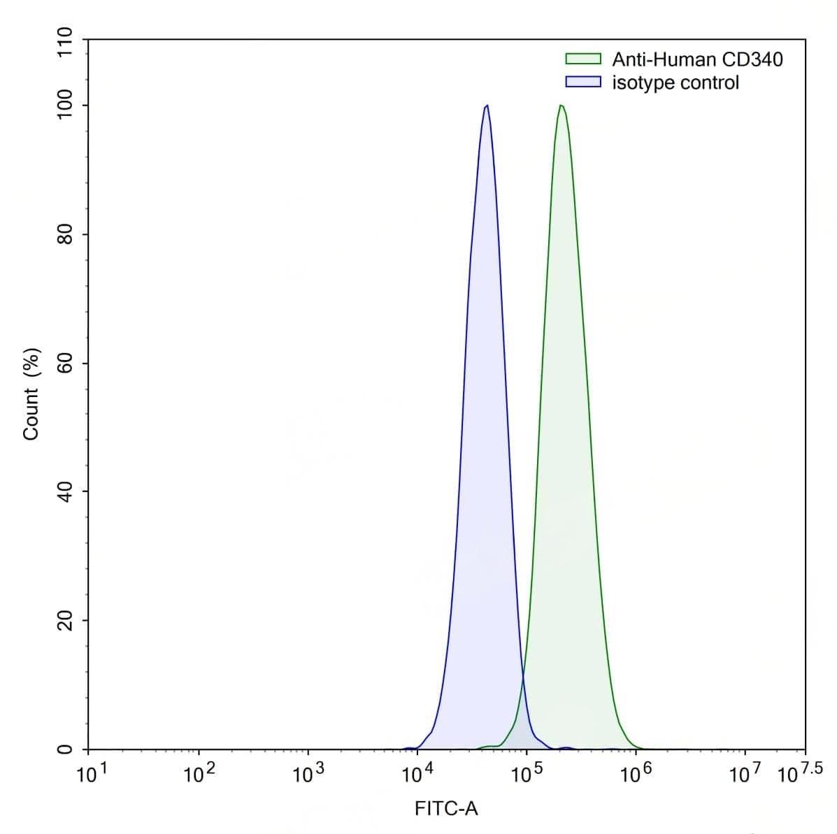
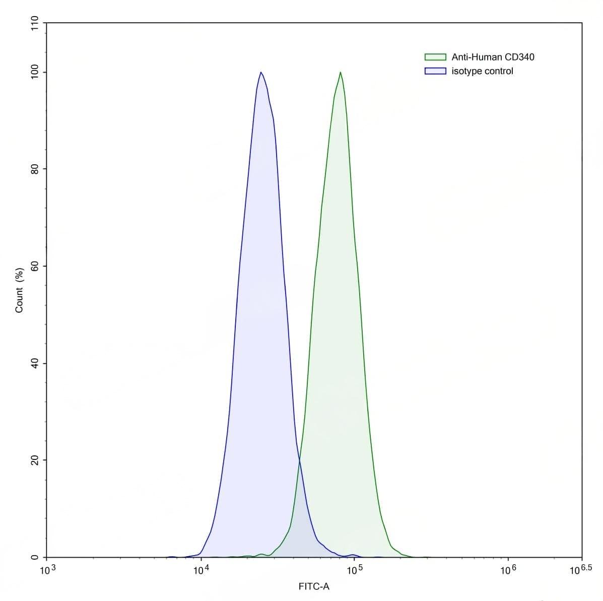
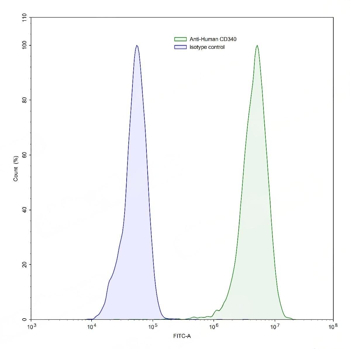
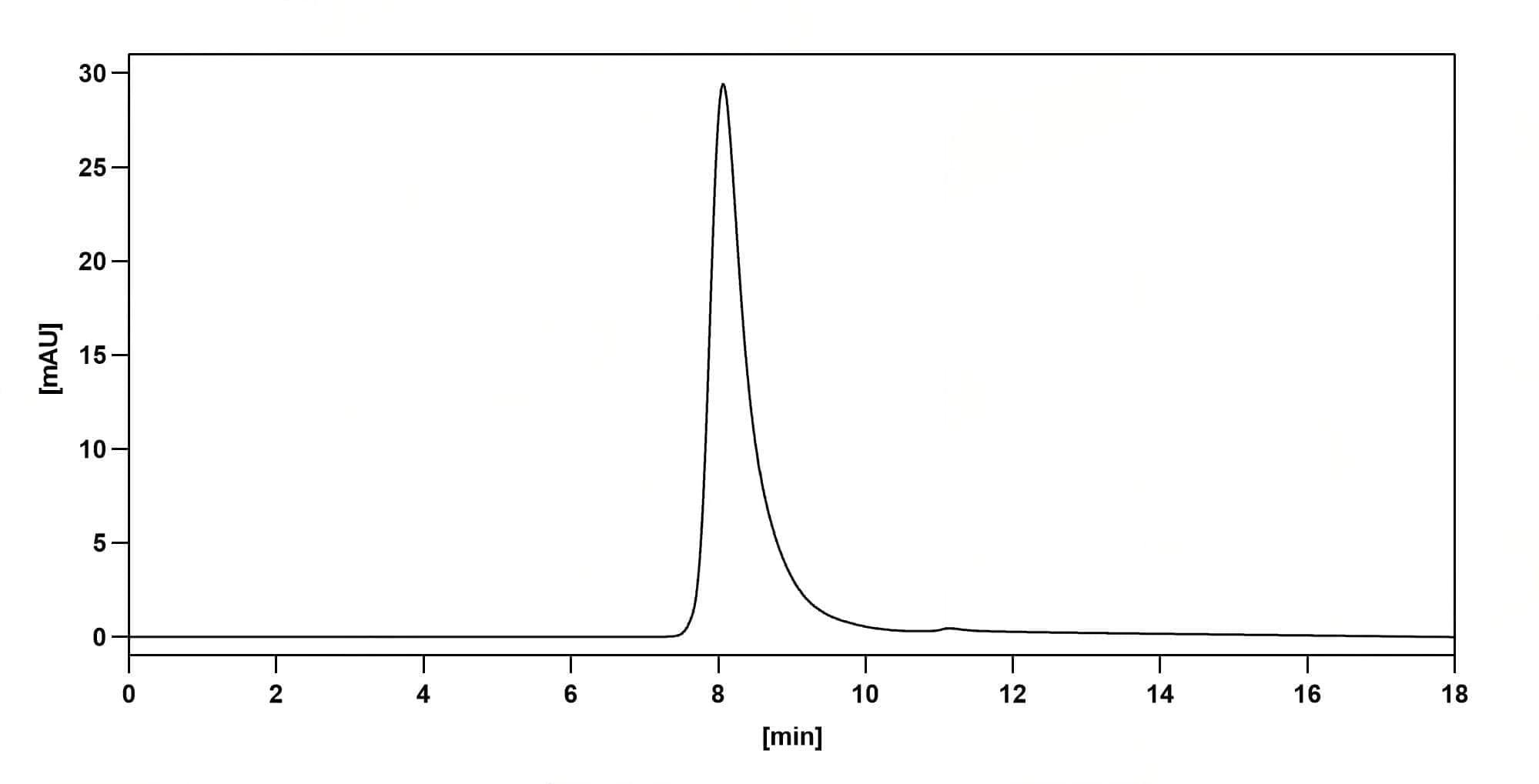
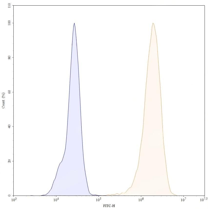
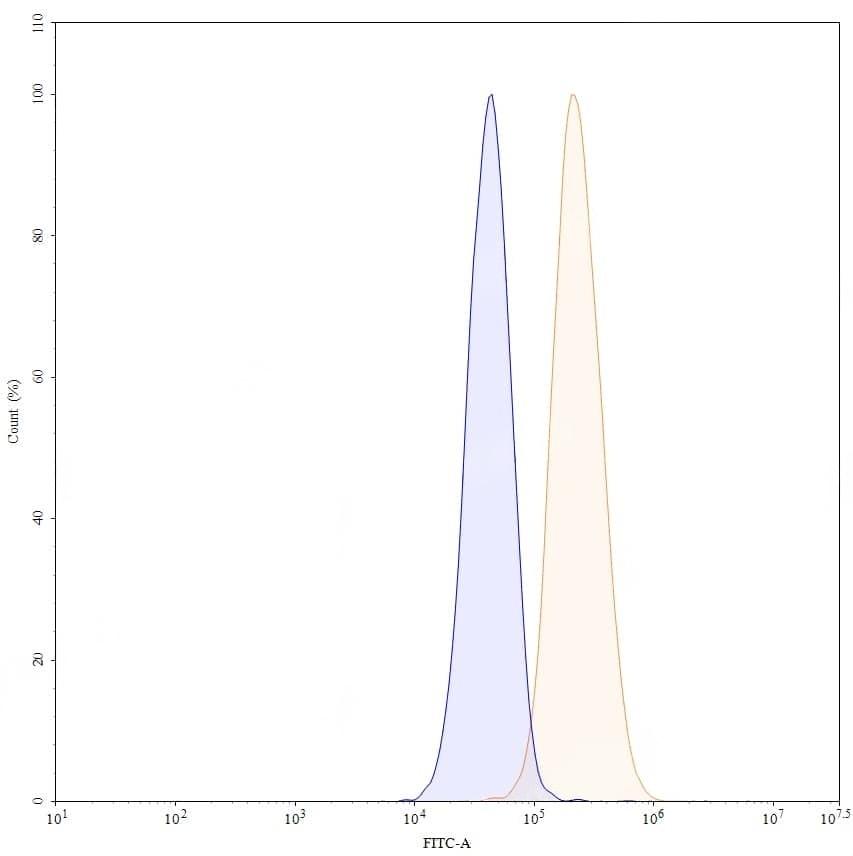
Flow-cytometry using anti-human CD340 antibody.MCF7 cells were stained with an irrelevant antibody (Blue Histogram) or an anti-human CD340 antibody monoclonal antibody (Catalog No.:TD-HY286107 ,Green Histogram) at a concentration of 5 ug/ml for 30 mins at RT. After washing, bound antibody was detected using a FITC conjugated goat anti-human antibody (Catalog No.:TD-HF690414) and cells analysed on a NovoCyte Flow Cytometer. | Flow-cytometry using anti-human CD340 antibody.MDA-MB-231 cells were stained with an irrelevant antibody (Blue Histogram) or an anti-human CD340 antibody monoclonal antibody (Catalog No.:TD-HY286107 ,Green Histogram) at a concentration of 5 ug/ml for 30 mins at RT. After washing, bound antibody was detected using a FITC conjugated goat anti-human antibody (Catalog No.:TD-HF690414) and cells analysed on a NovoCyte Flow Cytometer. | Flow-cytometry using anti-human CD340 antibody.SK-BR-3 cells were stained with an irrelevant antibody (Blue Histogram) or an anti-human CD340 antibody monoclonal antibody (Catalog No.:TD-HY286107 ,Green Histogram) at a concentration of 5 ug/ml for 30 mins at RT. After washing, bound antibody was detected using a FITC conjugated goat anti-human antibody (Catalog No.:TD-HF690414) and cells analysed on a NovoCyte Flow Cytometer. | SEC-HPLC detection for Anti-Human CD340/ERBB2/HER2 Antibody (4D5V8). | Flow-cytometry using anti-human CD340 antibody. SK-BR-3 cells were stained with an irrelevant antibody (Blue Histogram) or an anti-human CD340 monoclonal antibody (Catalog No.:TD-HY286107, Yellow Histogram) at a concentration of 5 µg/ml for 30 mins at RT. After washing, bound antibody was detected using a a Goat Anti-Human IgG H&L Polyclonal Antibody, FITC (Catalog No.:TD-HF690414) and cells analysed on a NovoCyte Flow Cytometer. | Flow-cytometry using anti-human CD340 antibody. MCF-7 cells were stained with an irrelevant antibody (Blue Histogram) or an anti-human CD340 monoclonal antibody (Catalog No.:TD-HY286107, Yellow Histogram) at a concentration of 5 µg/ml for 30 mins at RT. After washing, bound antibody was detected using a a Goat Anti-Human IgG H&L Polyclonal Antibody, FITC (Catalog No.:TD-HF690414) and cells analysed on a NovoCyte Flow Cytometer.