Protein A/G purified from cell culture supernatant.
Accession
P01375
Form
Liquid
Storage buffer
0.01M PBS, pH 7.4, 0.09% Sodium azide.Please refer to the specific buffer information in the hardcopy of datasheet or the lot-specific COA.
Stability and Storage
Use a manual defrost freezer and avoid repeated freeze-thaw cycles. Store at 4°C short term (1-2 weeks). Store at -20°C 12 months. Store at -80°C long term.
Caption
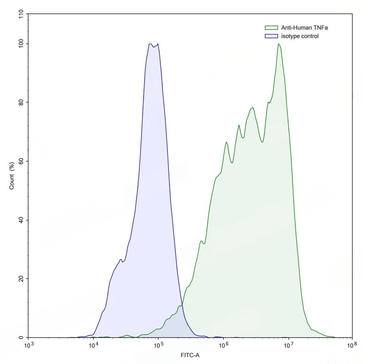
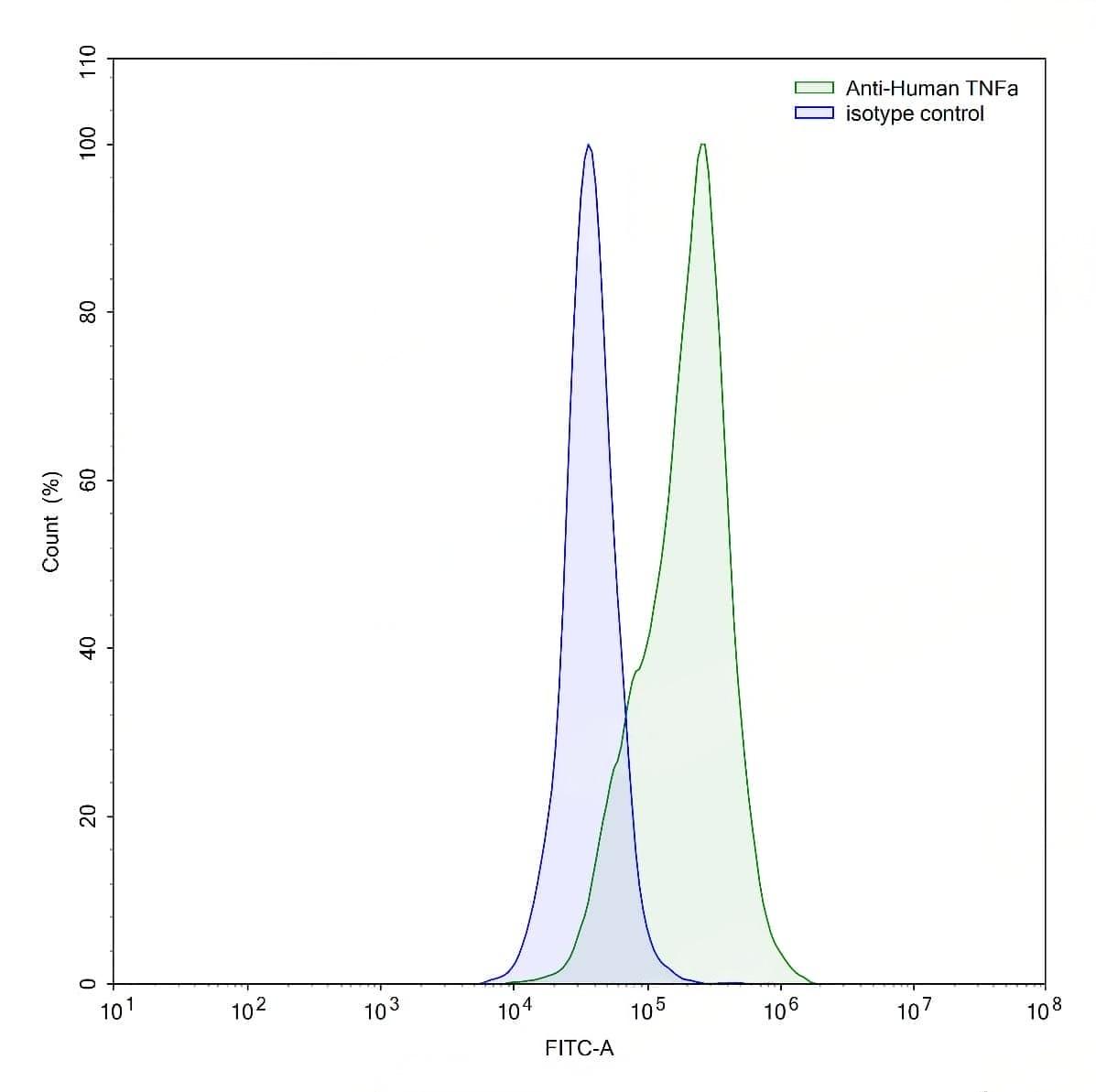
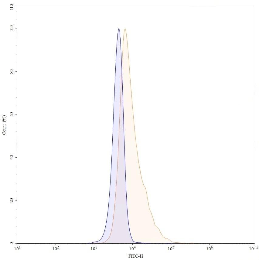
Flow-cytometry using anti-human TNFa antibody.TNFa Transfected CHO cells were stained with an irrelevant antibody (Blue Histogram) or an anti-human TNFa antibody monoclonal antibody (Catalog No.:TD-HF879107 ,Green Histogram) at a concentration of 5 ug/ml for 30 mins at RT. After washing, bound antibody was detected using a FITC conjugated goat anti-mouse antibody (Catalog No.:TD-MF690414) and cells analysed on a NovoCyte Flow Cytometer. | Flow-cytometry using anti-human TNFa antibody.LPS treated THP-1 cells were stained with an irrelevant antibody (Blue Histogram) or an anti-human TNFa antibody monoclonal antibody (Catalog No.:TD-HF879107 ,Green Histogram) at a concentration of 5 ug/ml for 30 mins at RT. After washing, bound antibody was detected using a FITC conjugated goat anti-mouse antibody (Catalog No.:TD-MF690414) and cells analysed on a NovoCyte Flow Cytometer. | Flow-cytometry using anti-human TNFa antibody. 293T transfected cells were stained with an irrelevant antibody (Blue Histogram) or an anti-human TNFa monoclonal antibody (Catalog No.:TD-HF879107, Yellow Histogram) at a concentration of 5 µg/ml for 30 mins at RT. After washing, bound antibody was detected using a Goat Anti-Mouse IgG H&L Polyclonal Antibody, FITC (Catalog No.:TD-MF690414) and cells analysed on a NovoCyte Flow Cytometer.