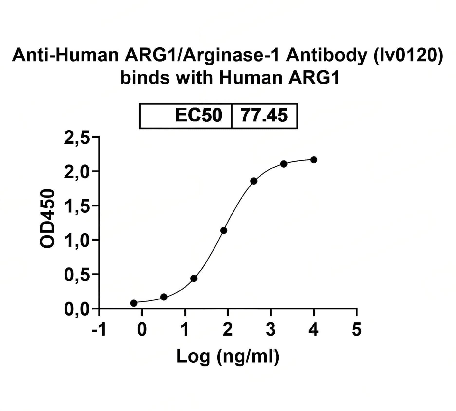
Caption
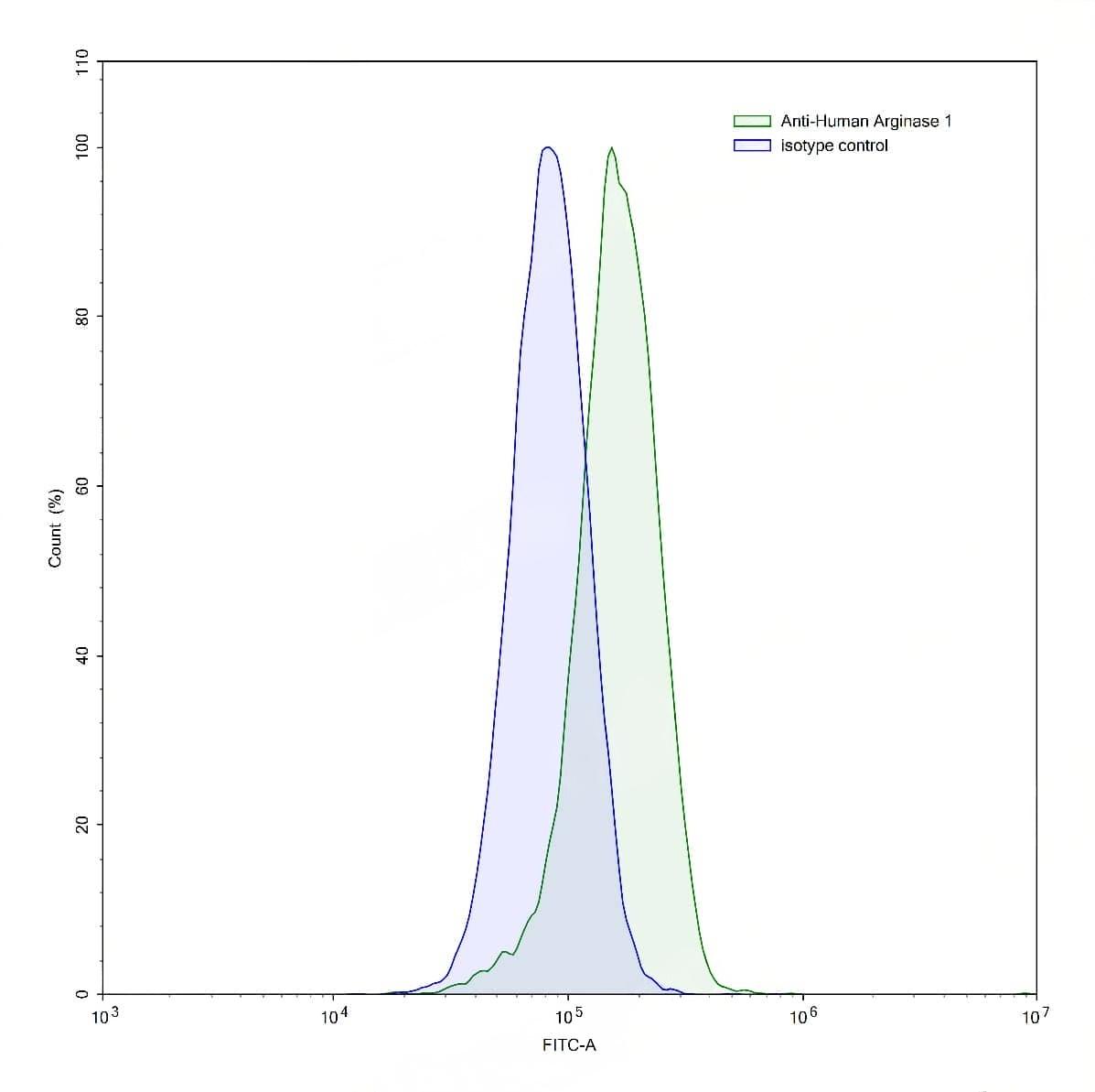
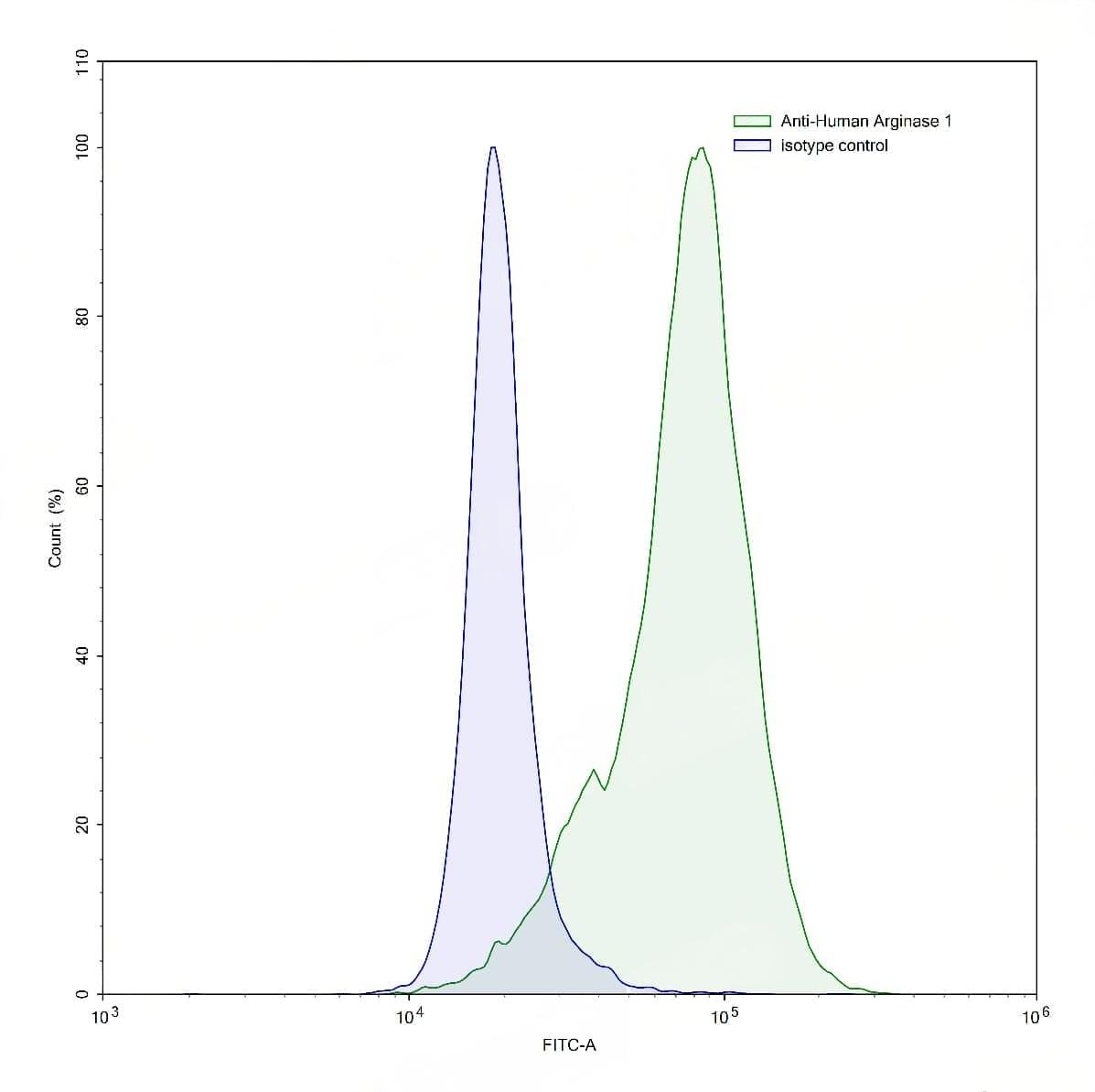
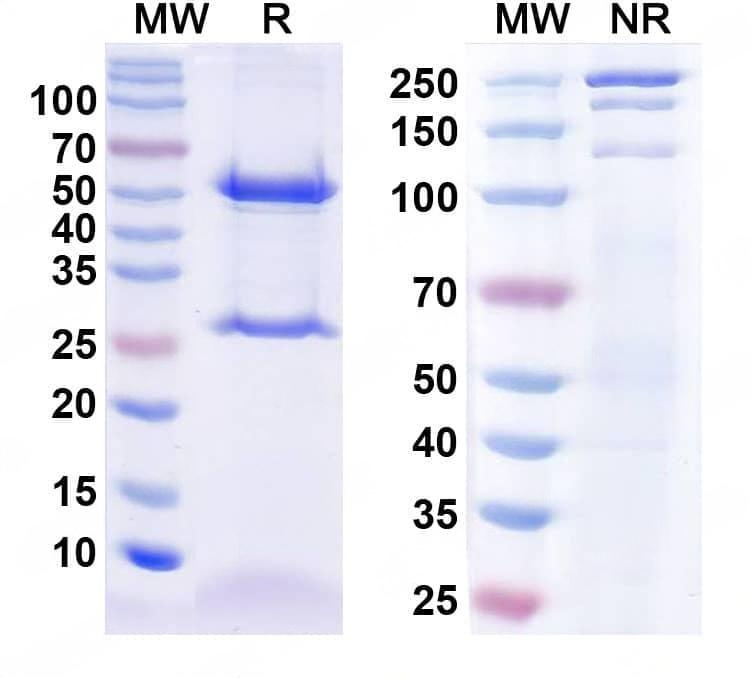
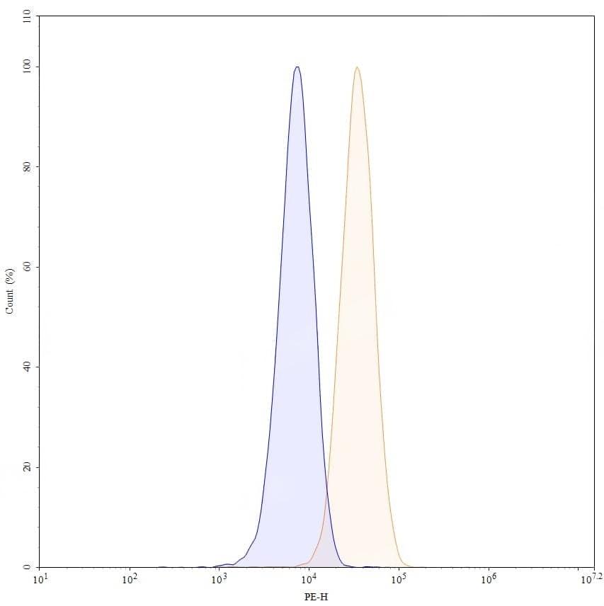
Detects Human ARG1/Arginase-1 in indirect ELISAs. | Flow-cytometry using anti-human Arginase 1 antibody.M2 macrophages derived from THP-1 cells were stained with an irrelevant antibody (Blue Histogram) or an anti-human Arginase 1 antibody monoclonal antibody (Catalog No.:TD-HY339010 ,Green Histogram) at a concentration of 5 µg/ml for 30 mins at RT. After washing, bound antibody was detected using a FITC conjugated goat anti-mouse antibody (Catalog No.:TD-MF690414) and cells analysed on a NovoCyte Flow Cytometer.,Flow-cytometry using anti-human Arginase 1 antibody.Human peripheral blood lymphocytes were stained with an irrelevant antibody (Blue Histogram) or an anti-human Arginase 1 antibody monoclonal antibody (Catalog No.:TD-HY339010 ,Green Histogram) at a concentration of 5 µg/ml for 30 mins at RT. After washing, bound antibody was detected using a FITC conjugated goat anti-mouse antibody (Catalog No.:TD-MF690414) and cells analysed on a NovoCyte Flow Cytometer. | SDS-PAGE for InVivoMAb Anti-Human ARG1/Arginase-1 Antibody (Iv0120). | Flow-cytometry using PE anti-human Arginase 1 antibody. HepG-2 cells were fixed and permeabilized, then stained with an irrelevant antibody (Blue Histogram) or an PE anti-human Arginase 1 monoclonal antibody (Catalog No.:TD-HY339010, Yellow Histogram) at a concentration of 5 µg/ml for 30 mins at RT. After washing, cells analysed on a NovoCyte Flow Cytometer.