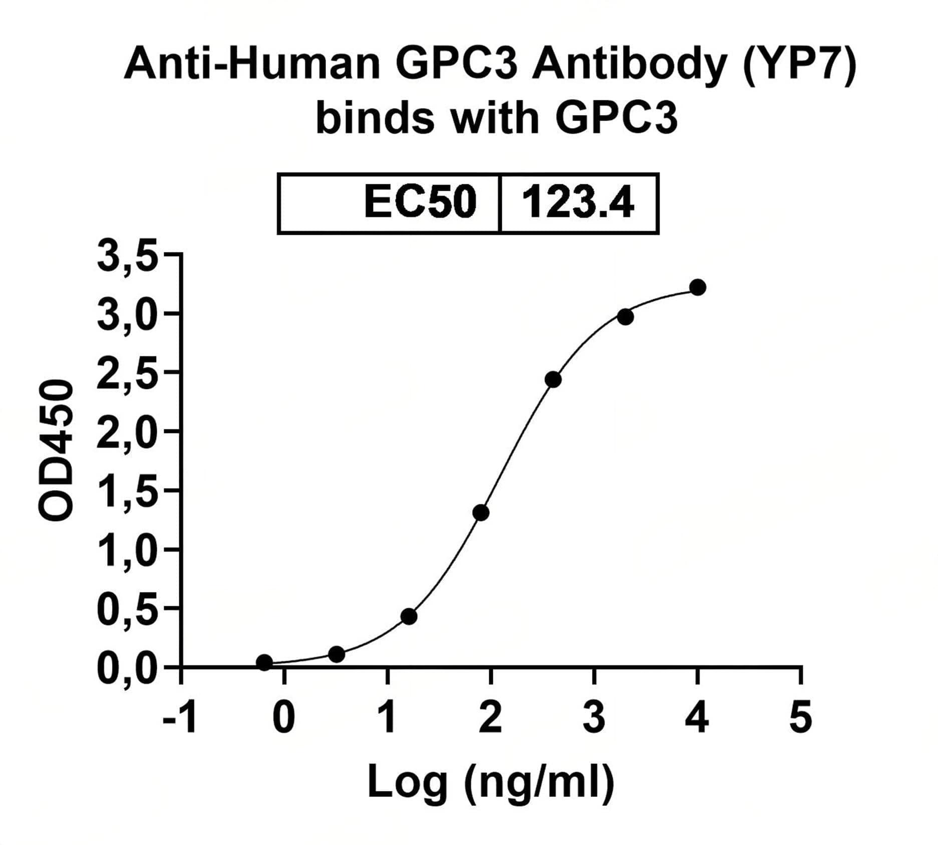
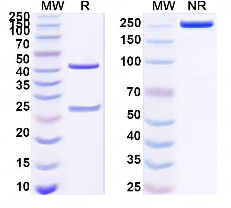
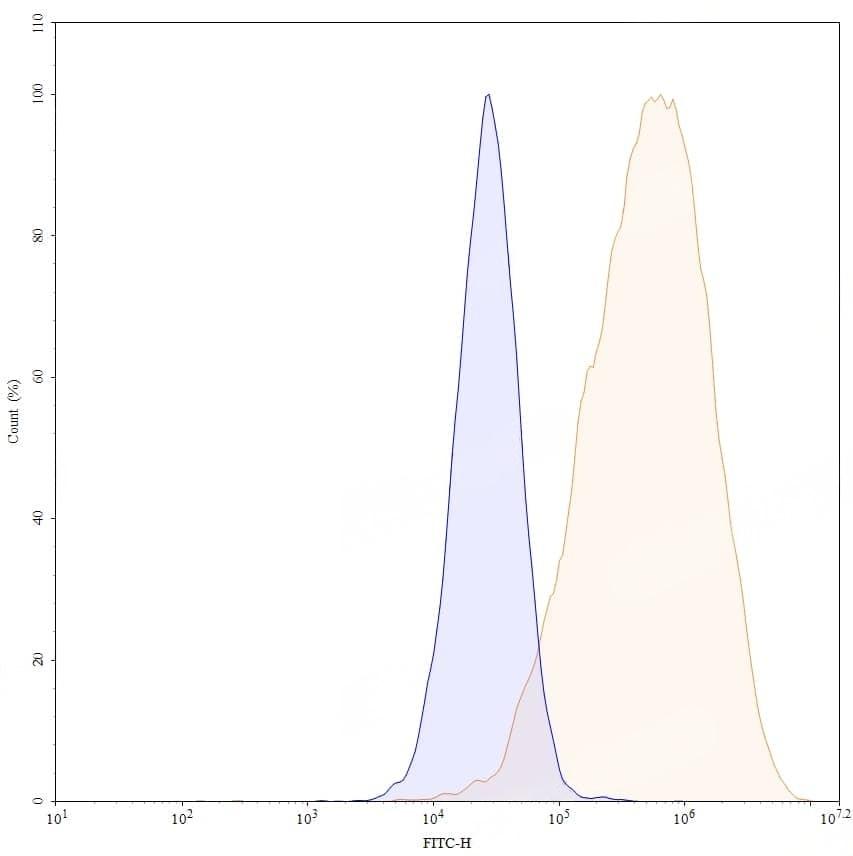
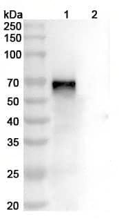
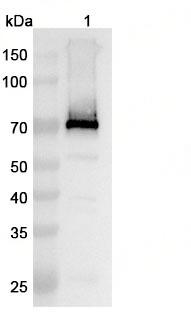
Caption
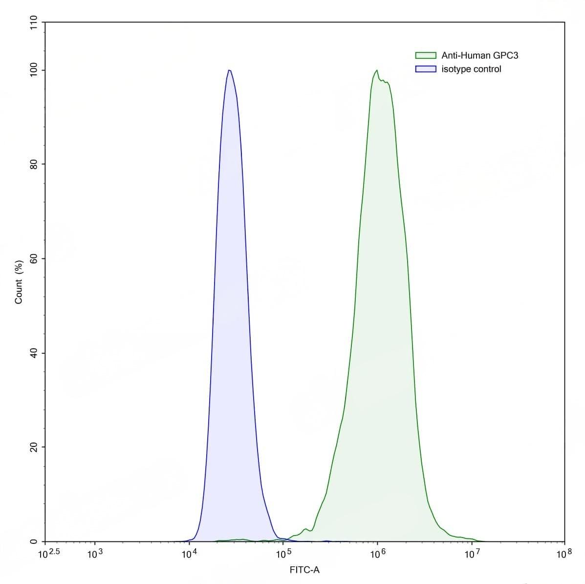
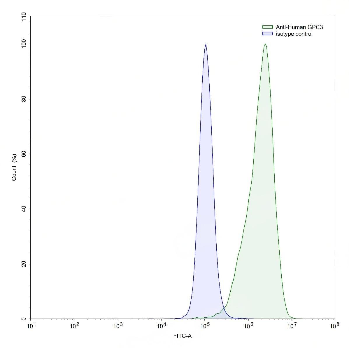
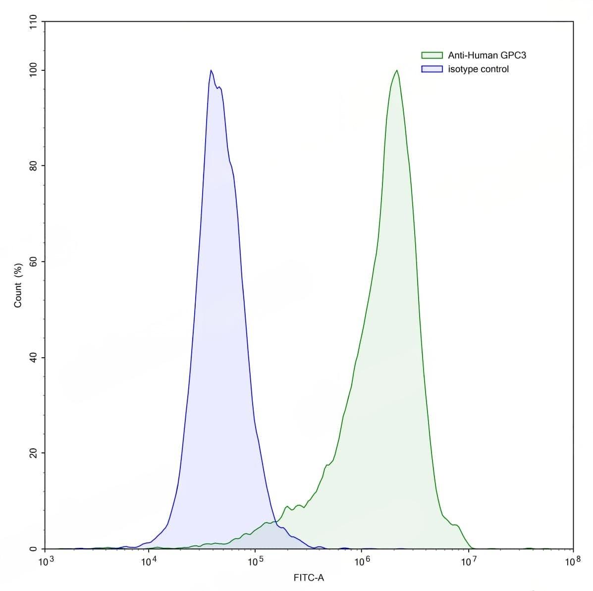
Detects Human GPC3 in indirect ELISAs. | Flow-cytometry using anti-human GPC3 antibody.GPC3 Transfected CHO cells were stained with an irrelevant antibody (Blue Histogram) or an anti-human GPC3 antibody monoclonal antibody (Catalog No.:TD-HW310023 ,Green Histogram) at a concentration of 5 µg/ml for 30 mins at RT. After washing, bound antibody was detected using a FITC conjugated goat anti-human antibody (Catalog No.:TD-HF690414) and cells analysed on a NovoCyte Flow Cytometer.,Flow-cytometry using anti-human GPC3 antibody.HepG2 cells were stained with an irrelevant antibody (Blue Histogram) or an anti-human GPC3 antibody monoclonal antibody (Catalog No.:TD-HW310023 ,Green Histogram) at a concentration of 5 µg/ml for 30 mins at RT. After washing, bound antibody was detected using a FITC conjugated goat anti-human antibody (Catalog No.:TD-HF690414) and cells analysed on a NovoCyte Flow Cytometer.,Flow-cytometry using anti-human GPC3 antibody.Human Jurkat cell line were stained with an irrelevant antibody (Blue Histogram) or an anti-human GPC3 antibody monoclonal antibody (Catalog No.:TD-HW310023 ,Green Histogram) at a concentration of 5 µg/ml for 30 mins at RT. After washing, bound antibody was detected using a FITC conjugated goat anti-human antibody (Catalog No.:TD-HF690414) and cells analysed on a NovoCyte Flow Cytometer. | SDS-PAGE for Anti-Human GPC3 Antibody (YP7). | Flow-cytometry using anti-human GPC3 antibody. Untransfected cells (blue Histogram) and Transfected cells (Yellow Histogram) were stained with an anti-human GPC3 monoclonal antibody (Catalog No.:TD-HW310023) at a concentration of 5 µg/ml for 30 mins at RT. After washing, bound antibody was detected using a Goat Anti-Human IgG H&L Polyclonal Antibody, FITC (Catalog No.:TD-HF690414) and cells analysed on a NovoCyte Flow Cytometer. | Western blot analysis was performed using anti-GPC3/Glypican 3 monoclonal antibody at 1µg/ml on various samples.Lane 1:recombinant mouse GPC3/Glypican 3 (Catalog No.:TD-MW310011)Lane 2:negative control,Western blot analysis was performed using anti-GPC3 monoclonal antibody at 1ug/mL on various samples.Lane 1:HepG2 cell lysate