Target
AICD-57, A4, Beta-CTF, Protease nexin-II, Gamma-CTF(50), ABPP, S-APP-beta, APP, Abeta40, Abeta42, AID(59), Amyloid intracellular domain 59, AID(57), S-APP-alpha, Gamma-CTF(59), Beta-secretase C-terminal fragment, Amyloid-beta precursor protein, Amyloid precursor protein, Beta-APP42, PreA4, Alzheimer disease amyloid protein, APPI, PN-II, AICD-59, Alpha-secretase C-terminal fragment, Amyloid-beta A4 protein, Amyloid intracellular domain 57, CVAP, Amyloid intracellular domain 50, Beta-APP40, AD1, Cerebral vascular amyloid peptide, AID(50), Alpha-CTF, Gamma-CTF(57), AICD-50
Caption
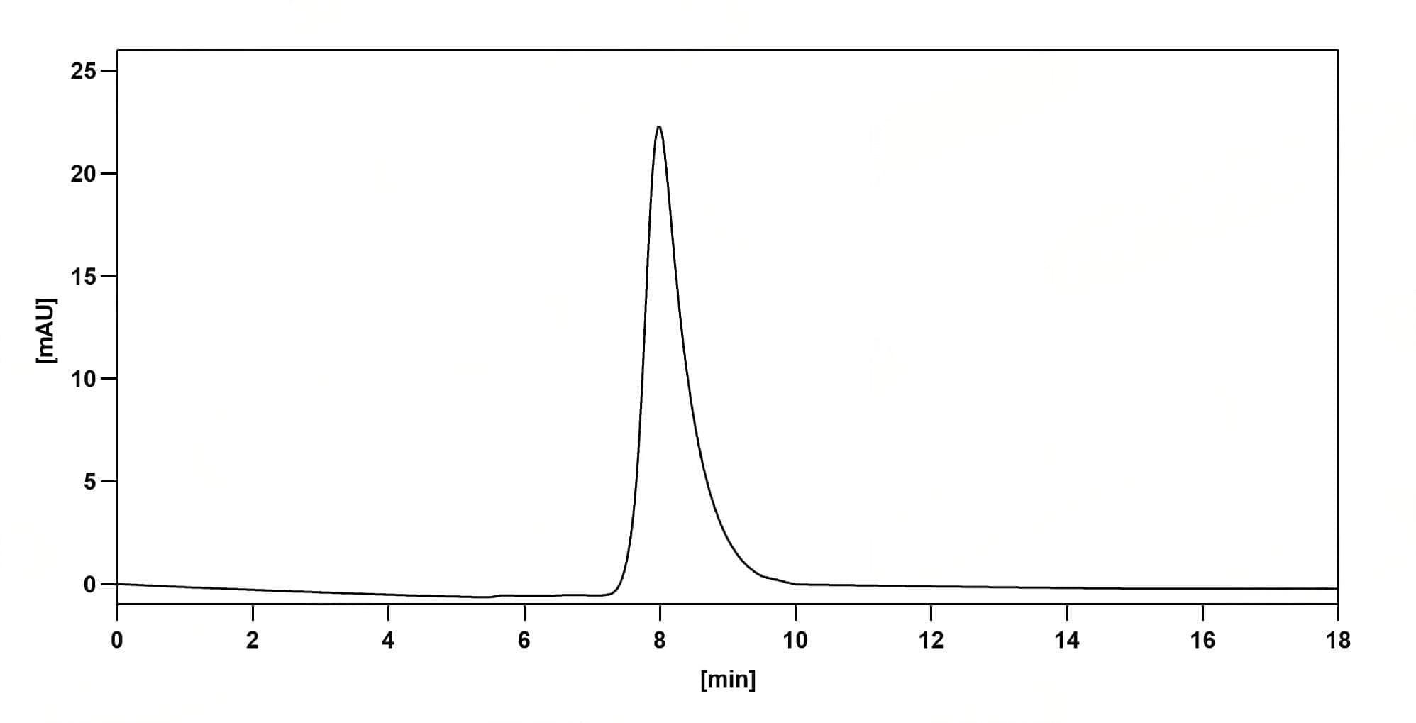
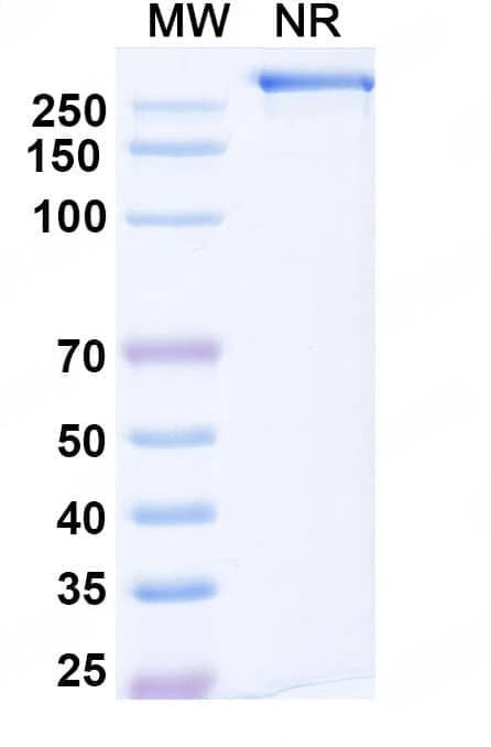
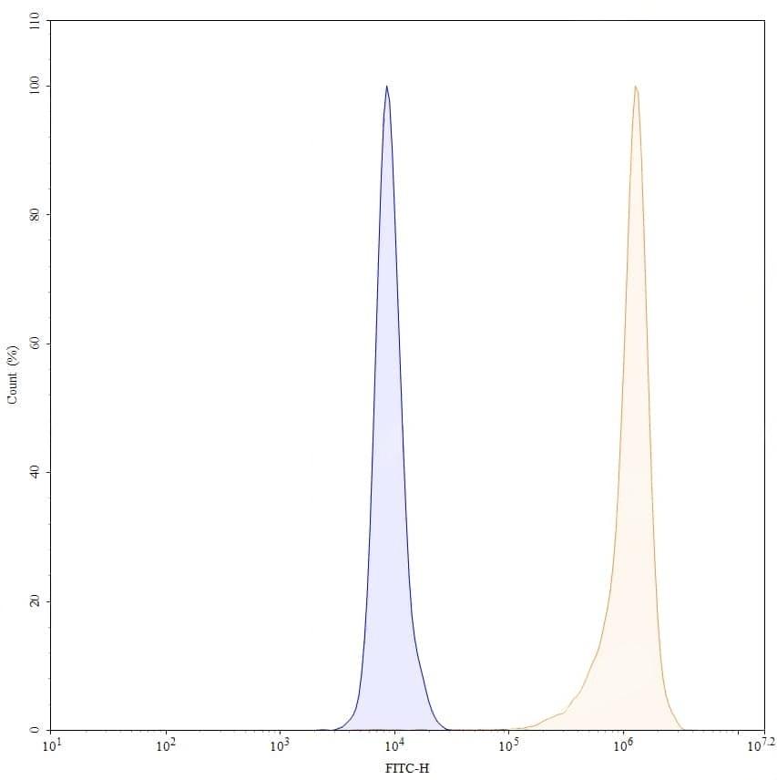
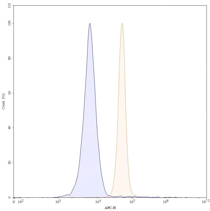
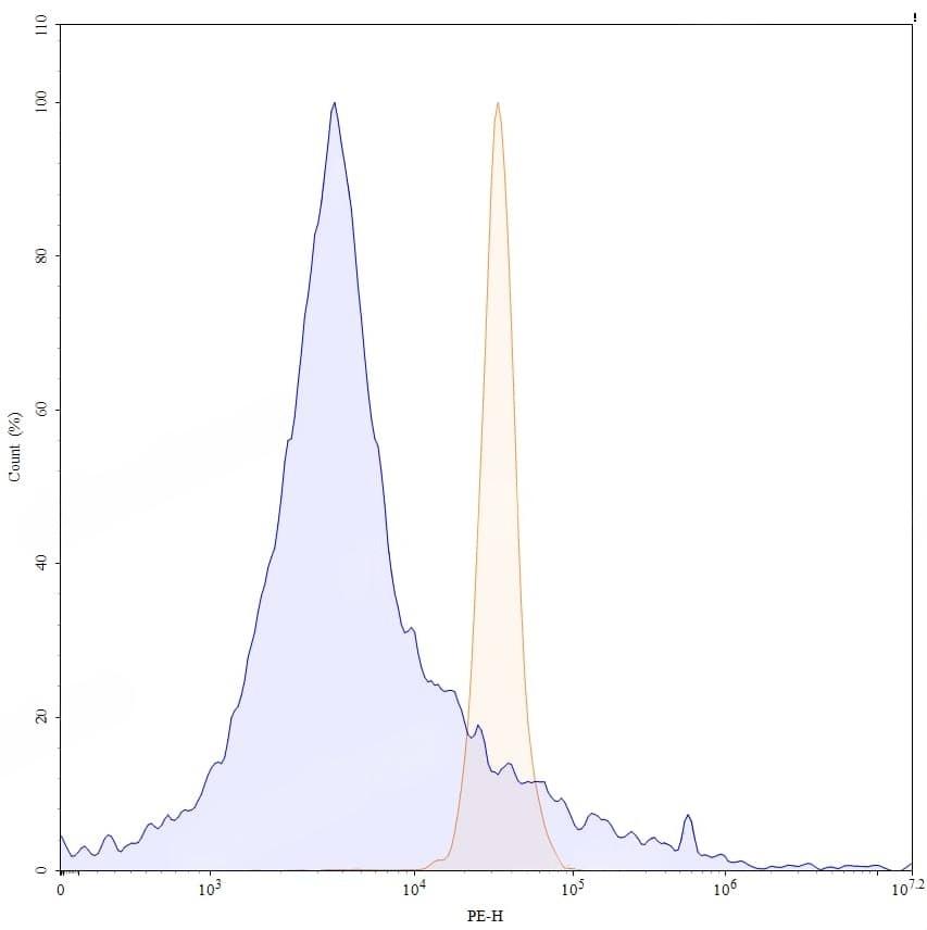
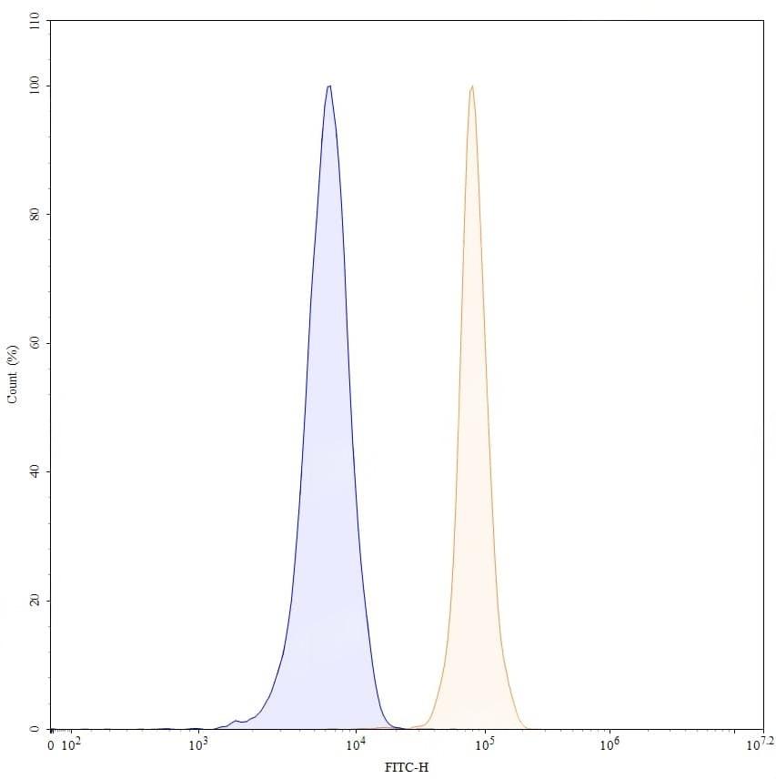
SEC-HPLC detection for Research Grade Gantenerumab. | SDS-PAGE for Research Grade Gantenerumab. | Flow-cytometry using anti-human APP antibody. SH-SY5Y cells were fixed and permeabilized, then stained with an irrelevant antibody (Blue Histogram) or an anti-human APP monoclonal antibody (Catalog HY235036, Yellow Histogram) at a concentration of 5 µg/ml for 30 mins at RT. After washing, bound antibody was detected using Goat Anti-Human IgG H&L Polyclonal Antibody, FITC (abinScience: HF690414) and cells analysed on a NovoCyte Flow Cytometer. | Flow-cytometry using APC anti-human APP antibody. SH-SY5Y cells were fixed and permeabilized, then stained with an irrelevant antibody (Blue Histogram) or an APC anti-human APP monoclonal antibody (Catalog HY235036, Yellow Histogram) at a concentration of 5 µg/ml for 30 mins at RT. After washing, cells analysed on a NovoCyte Flow Cytometer. ### Flow-cytometry using PE anti-human APP antibody. SH-SY5Y cells were fixed and permeabilized, then stained with an irrelevant antibody (Blue Histogram) or an PE anti-human APP monoclonal antibody (Catalog HY235036, Yellow Histogram) at a concentration of 5 µg/ml for 30 mins at RT. After washing, cells analysed on a NovoCyte Flow Cytometer. ### Flow-cytometry using FITC anti-human APP antibody. SH-SY5Y cells were fixed and permeabilized, then stained with an irrelevant antibody (Blue Histogram) or an FITC anti-human APP monoclonal antibody (Catalog HY235036, Yellow Histogram) at a concentration of 5 µg/ml for 30 mins at RT. After washing, cells analysed on a NovoCyte Flow Cytometer.